Engineering · Explant analysis
Explant analysis
The examination of a device removed from a patient, with or without a complication: failure characterisation, device degradation and clinical feedback.
Our approach
Every explanted device is a data point
Our goal is not solely to assess vigilance cases, but to offer a unique service that studies and categorises the defects that can affect implantable medical devices, across every surgical specialty.
Every explanted device, whether or not it is associated with a complication, is analysed within a European network of surgeons. Over the years we have built one of the largest explant databases in the world.
1 800+
explanted implantable medical devices analysed since Gepromed was founded
150+
vascular explants received in 2023
≈ 290
scientific publications informed by this work
Five pillars
What our expertise rests on
01
Patient safety
Better understanding how materials age, to improve patient care.
02
Vigilance
Objective, robust data for the authorities.
03
Manufacturer support
Helping with root-cause analysis and the continuous improvement of devices.
04
Multidisciplinary expertise
A team of clinical experts and textile experts.
05
A complete platform
From macroscopic to microscopic analysis, by way of radiography.
Which explants
Vascular first, and beyond
Our reference field
Vascular explants
The field in which we are recognised as experts today.
Also
Abdominal wall reinforcement and gynaecology
Our experience also extends to abdominal wall reinforcement devices and gynaecological devices.
Custom
Other devices
Beyond these fields, we can build with our experts a project tailored to your needs.
The protocol
From reception to report
Our analysis protocol has been ISO 9001 certified since 6 December 2012.
01
Logging
Each explant is logged on reception.
02
Macroscopic analysis
A first examination of the device, before cleaning.
03
Complementary analyses
On request: radiography, histology, macroscopic and microscopic studies after enzymatic cleaning.
04
Detailed report
All the findings, brought together in a report.
The equipment
From macroscopic to microscopic
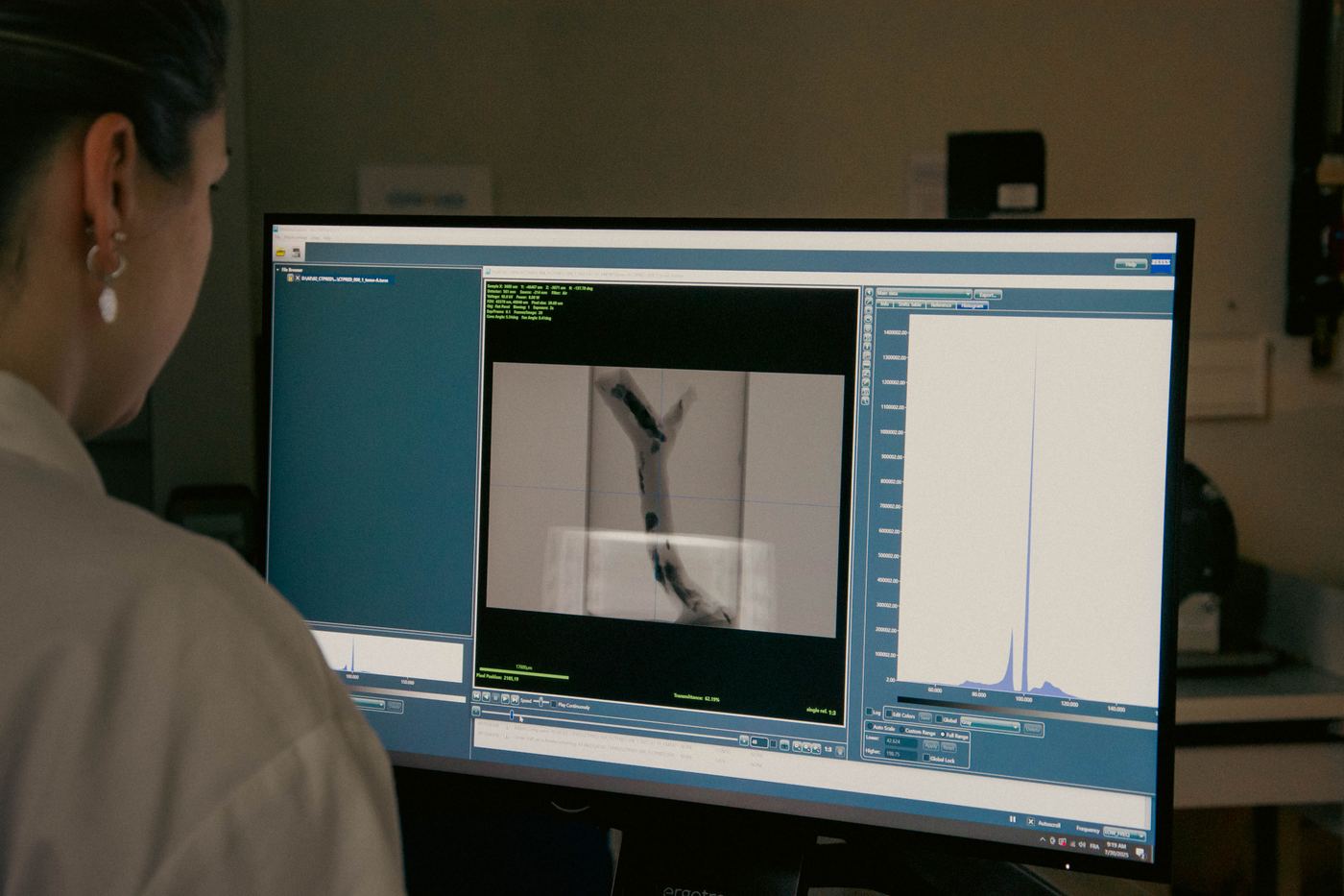
Radiography, tomography, microscopy, surface analysis and histology, on a single platform.
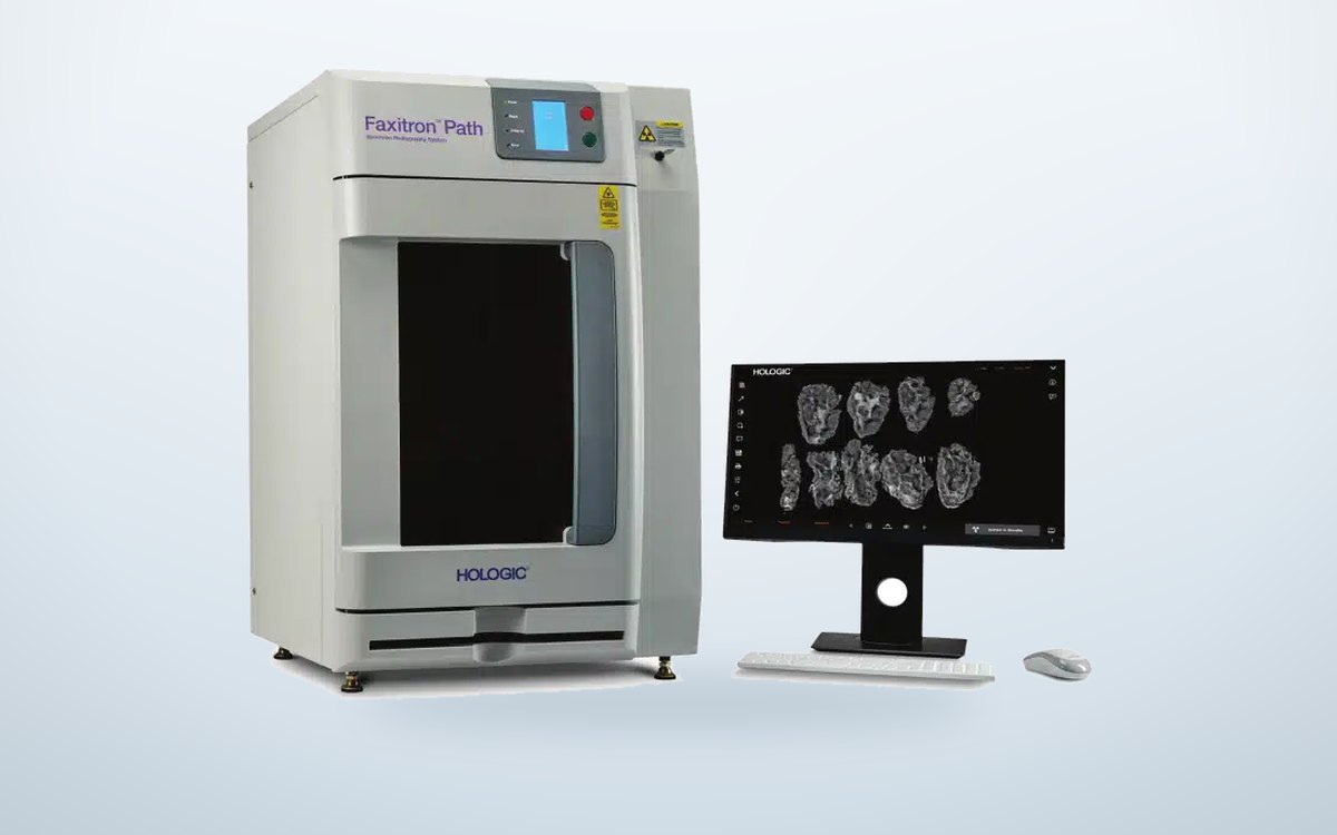 Radiography
Faxitron PathVision
High-resolution radiography for detailed imaging of samples.
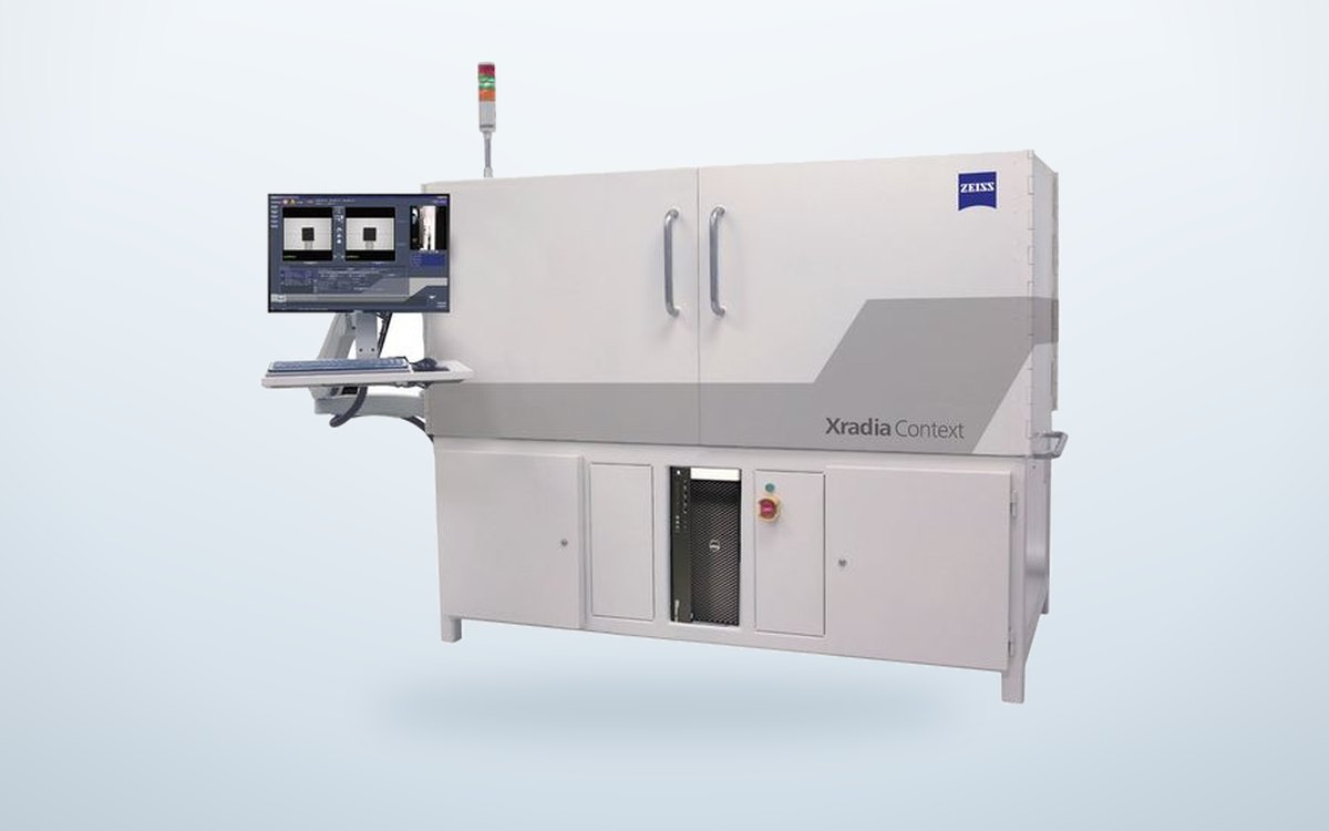 Micro-CT
ZEISS Xradia Context
Tomography and internal volumetric 3D imaging.
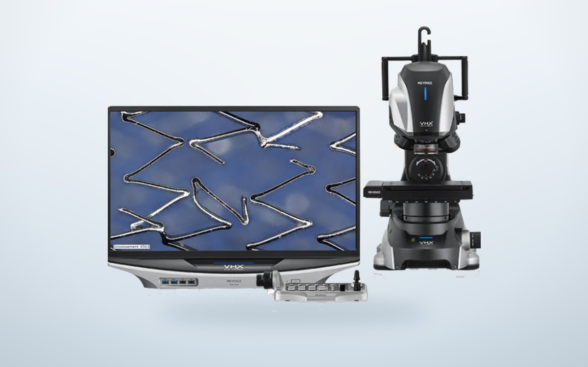 3D imaging
Keyence VHX-7100
High-resolution 3D reconstruction, from ×20 to ×2500.
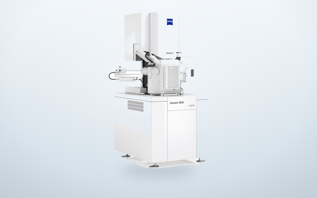 Microscopy
ZEISS SEM 300
Scanning electron microscopy for morphological analysis.
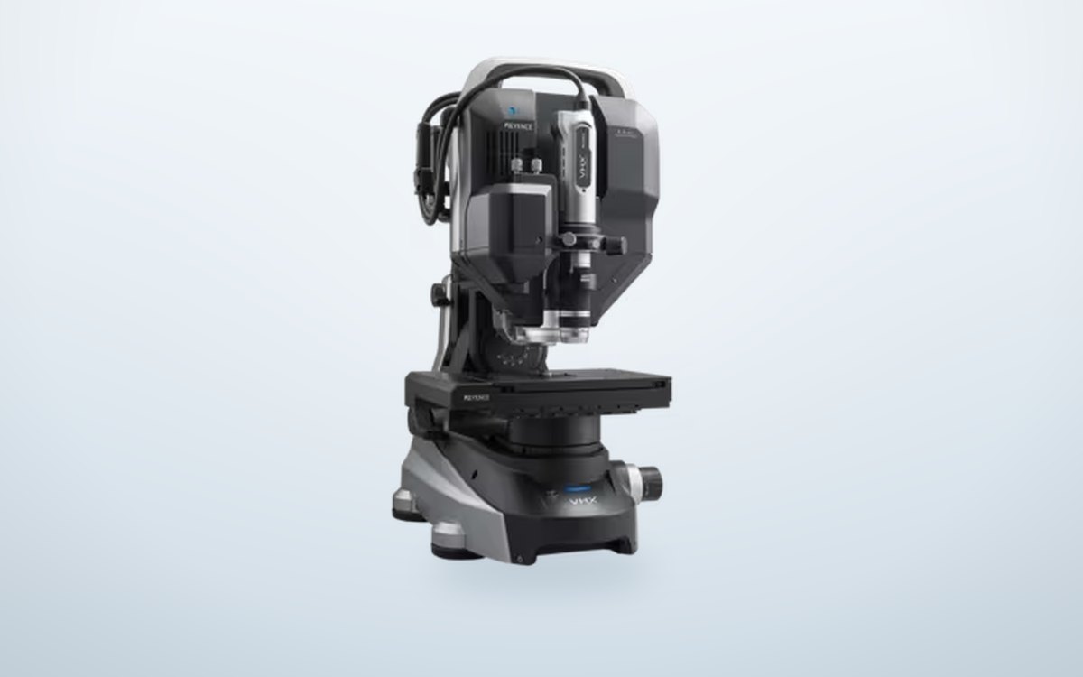 Surface
Keyence VHX-S7000E + EA-300
Analysis of surface characteristics.
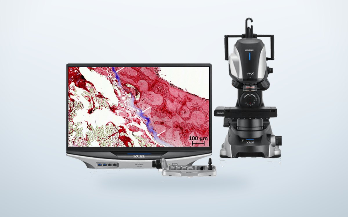 Histology
Histology
Histological analysis of biological and tissue sections.
These instruments, except the micro-CT, can also be booked for your own campaigns: see the Equipment rental page →
Send us an explant
A bottle, a form, an address
- Place the sample in a sealed bottle containing 10% formalin (approx. 4% formaldehyde).
- Enclose the clinical data sheet: fiche de recueil (FR, PDF) · download the clinical data sheet (EN, PDF)
- Ship it to Gepromed:
Bâtiment d'anesthésiologie
4 rue Kirschleger, 67085 Strasbourg Cedex, France
Out of transport kits? Email explant@gepromed.com with your work postal address: we will send you bottles and data sheets.
Beyond explants
Have another sample analysed
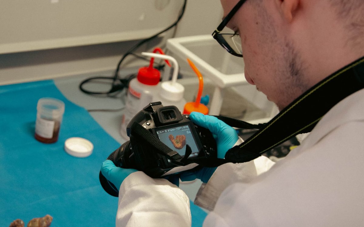
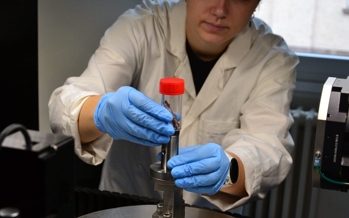
Optical, radiographic and tomographic: our imaging equipment can adapt to medical devices, to industrial materials or to research projects from other fields, as long as our technologies meet the needs of the analysis.
FAQ
Frequently asked questions
The examination of a device removed from a patient, with or without a complication. The analysis covers failure characterisation, device degradation and clinical feedback. It relies on radiographic and tomographic imaging, microscopy, surface analysis and histology.
No. Every explanted device is analysed, whether or not it is associated with a complication.
We are recognised today as experts in vascular explants, but our experience also extends to abdominal wall reinforcement devices and gynaecological devices. Beyond these fields, we can build with our experts a custom project tailored to your specific needs.
Yes, that is entirely possible, provided our technologies meet the needs of the sample's analysis.
Entrust us with an explant
An explanted device to analyse
- What we analyse
- Any explanted device, whether or not associated with a complication.
- What you receive
- A report with photographs and observations.
- Turnaround
- 10 days from receipt of the explant.
No-obligation request. Our team will get back to you to confirm availability and prepare a quote.